How Does Breast Cancer Look Like In Ultrasound : A A Simple Breast Cyst 75 B Breast Ultrasound Showing A Cancer Download Scientific Diagram / This high amount of echo results in a bright white spot appearing on the ultrasound image.
Dapatkan link
Facebook
X
Pinterest
Email
Aplikasi Lainnya
How Does Breast Cancer Look Like In Ultrasound : A A Simple Breast Cyst 75 B Breast Ultrasound Showing A Cancer Download Scientific Diagram / This high amount of echo results in a bright white spot appearing on the ultrasound image.. If your breast tissue is too dense for a mammogram. What does breast cancer look like on a mammogram? Screening mammograms have been used since the 1980s. Ultrasound is not used on its own as a screening test for breast cancer. A breast ultrasound is most often done to find out if a problem found by a mammogram or physical exam of the breast may be a cyst filled with fluid or a solid tumor.
Ultrasound imaging allows better evaluation of the status of the axillary lymph nodes in patients with ibc, an important step in determining extent of disease prior to initiation of chemotherapy. However, a dark spot on your ultrasound doesn't mean that you. It is the most common cause of cancer death in women. in 2005 alone, 519 000 deaths were recorded due to breast cancer. this means that one in every 100 deaths worldwide and almost one in every 15 cancer deaths were due to breast cancer. On ultrasound, a breast cancer tumor is often seen as hypoechoic, has irregular borders, and may appear spiculated. In the table the differences in ultrasound appearances are listed.
Molecular Markers Pathology And Ultrasound Features Of Invasive Breast Cancer Clinical Imaging from els-jbs-prod-cdn.jbs.elsevierhealth.com Sometimes breast cancer can look like a fibroadenoma and fibroadenomas can look like a cancer on ultrasound. What does the doctor look for on a mammogram? cancer.org. You can get dressed straight after the ultrasound. On ultrasound, a breast cancer tumor is often seen as hypoechoic, has irregular borders, and may appear spiculated. When is breast ultrasound used? Benign and malignant characteristics of breast lesions at ultrasound allow the classification as either malignant, intermediate or benign based on work published by stavros et al. Ultrasound characterization of breast masses. indian journal of radiology and imaging. However, a dark spot on your ultrasound doesn't mean that you.
Ultrasound is not used on its own as a screening test for breast cancer.
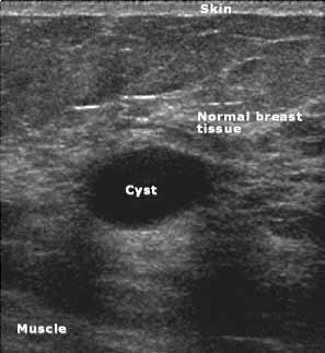
Cysts, tumors, and growths will appear as dark areas on the scan. Breast ultrasound uses sound waves to make a computer picture of the inside of the breast. What does a solid mass look like in an ultrasound breast image? Ultrasound of triple negative breast cancer. This is a network of thin tubes (vessels) and nodes that carry a clear fluid called lymph around the body. A breast ultrasound is most often done to find out if a problem found by a mammogram or physical exam of the breast may be a cyst filled with fluid or a solid tumor. Solid lesions can be a little brighter or darker than the surrounding tissue, and the way to evaluate these on ultrasound is to look closely at the margins or the outer edges of the nodule. In the table the differences in ultrasound appearances are listed. Ibc is a rapidly progressive, aggressive form of breast cancer associated with a low overall survival rate. On the flip side, there are benign (not cancerous) breast changes which can mimic breast cancer as well. What does breast cancer look like on a mammogram? American cancer society, 9 oct 2017. Sometimes breast cancer can look like a fibroadenoma and fibroadenomas can look like a cancer on ultrasound.
Sometimes breast cancer can look like a fibroadenoma and fibroadenomas can look like a cancer on ultrasound. Ultrasound imaging allows better evaluation of the status of the axillary lymph nodes in patients with ibc, an important step in determining extent of disease prior to initiation of chemotherapy. Any area that does not look like normal tissue is a possible cause for concern. However, a dark spot on your ultrasound doesn't mean that you. If you're younger than 25.
Breast Biopsy Ultrasound Guided from www.radiologyinfo.org However, a dark spot on your ultrasound doesn't mean that you. It is the most common cause of cancer death in women. in 2005 alone, 519 000 deaths were recorded due to breast cancer. this means that one in every 100 deaths worldwide and almost one in every 15 cancer deaths were due to breast cancer. Other ultrasound findings that suggest breast cancer include: A breast ultrasound is most often done to find out if a problem found by a mammogram or physical exam of the breast may be a cyst filled with fluid or a solid tumor. If a solid lump shows on the scan you might need to have. You might not need any further tests if everything looks normal. Tumor size is an important factor in breast cancer staging, and it can affect a person's treatment options and outlook. This high amount of echo results in a bright white spot appearing on the ultrasound image.
This is because it may miss some early signs of cancer.
This breast cancer ultrasound image shows changes related to breast cancer that are not seen as microcalcifications or a mass or lump. Any area that does not look like normal tissue is a possible cause for concern. If your breast tissue is too dense for a mammogram. Ultrasound imaging allows better evaluation of the status of the axillary lymph nodes in patients with ibc, an important step in determining extent of disease prior to initiation of chemotherapy. If a solid lump shows on the scan you might need to have. It is the most common cause of cancer death in women. in 2005 alone, 519 000 deaths were recorded due to breast cancer. this means that one in every 100 deaths worldwide and almost one in every 15 cancer deaths were due to breast cancer. With ultrasound, the radiologist will probably be trying to get a sense of the internal texture of the breast lesion and surrounding area. Physical examination and mammogram can be more accurate in some settings. If there are calcifications within the nodular dcis, one may be able to see these on ultrasound as white flecks. The images that a breast ultrasound produces are in black and white. A diagnostic mammogram is used to check for breast cancer when there is a sign or symptom of disease. A breast ultrasound is most often done to find out if a problem found by a mammogram or physical exam of the breast may be a cyst filled with fluid or a solid tumor. To look more closely at a.
Any area that does not look like normal tissue is a possible cause for concern. Below are images of dcis on breast ultrasound. Ultrasound is not used on its own as a screening test for breast cancer. Ultrasound (us) shows an irregular, hypoechoic (dark gray) spiculated mass (arrow), highly suspicious for cancer. This is because it may miss some early signs of cancer.
Staging Of Breast Cancer With Ultrasound Sciencedirect from ars.els-cdn.com Ultrasound characterization of breast masses. indian journal of radiology and imaging. Other names for this test: Ultrasound is only one means of evaluation of the breast. Tumors are likely to be smaller when doctors detect them early, which can. Breast cancer is among the most common causes of cancer deaths today, coming fifth after lung, stomach, liver and colon cancers. Any area that does not look like normal tissue is a possible cause for concern. In the table the differences in ultrasound appearances are listed. Overall, 57 (20.8%) of the 274 women had cancer in the axillary lymph nodes.
A diagnostic mammogram is used to check for breast cancer when there is a sign or symptom of disease.
Other names for this test: Sometimes breast cancer can look like a fibroadenoma and fibroadenomas can look like a cancer on ultrasound. Breast cancer is among the most common causes of cancer deaths today, coming fifth after lung, stomach, liver and colon cancers. If your breast tissue is too dense for a mammogram. Doctors often use them to guide a needle during a biopsy. Rather, the right breast is seen as smaller than the left breast. Sometimes the cancer cells can spread into the nearby lymph nodes. There is a slight increase in the density in the right breast compared with the left. What does the doctor look for on a mammogram? cancer.org. The images that a breast ultrasound produces are in black and white. You might not need any further tests if everything looks normal. A screening mammogram is performed at regular intervals to check for breast cancer in women who have no signs or symptoms of the disease. If a solid lump shows on the scan you might need to have.
Dinamai asy syams (matahari) diambil dari perkataan asy syams yang terdapat pada ayat permulaan surat ini. Dan kaumnya dalam surat ini. Surat ini dinamai surat huud karena ada hubungan dengan terdapatnya kisah nabi huud a.s. ۞ وَمَا مِنْ دَابَّةٍ فِي الْأَرْضِ إِلَّا عَلَى اللَّهِ رِزْقُهَا وَيَعْلَمُ مُسْتَقَرَّهَا وَمُسْتَوْدَعَهَا ۚ كُلٌّ فِي كِتَابٍ مُبِينٍ. Surah ini diturunkan di makkah sehingga tergolong surah makiyah dan terdiri dari tujuh ayat. Quran: 11. Surat Hud (Prophet Hud): Arabic and English from i.ytimg.com Dan kaumnya dalam surat ini. ۞ وَمَا مِنْ دَابَّةٍ فِي الْأَرْضِ إِلَّا عَلَى اللَّهِ رِزْقُهَا وَيَعْلَمُ مُسْتَقَرَّهَا وَمُسْتَوْدَعَهَا ۚ كُلٌّ فِي كِتَابٍ مُبِينٍ. Surat ini dinamai surat huud karena ada hubungan dengan terdapatnya kisah nabi huud a.s. Surah ini diturunkan di makkah sehingga tergolong surah makiyah...
Best Software For Making Cards : Greeting Card Factory Deluxe 11 Upgrade | Greeting Card ... - You can save created game cards as pdf and jpg files. . Card maker is a simple game card maker software for windows. It also features qr codes; A beautiful greeting card, wedding invitation, poster. Photo collage software is simple. Card making templates for microsoft word while most people associate microsoft word with basic word processing functions, this software can actually be used to design your own greeting cards. The potential design combinations are infinite when you make your card with adobe spark. The software is mainly known to create various numbers of id cards and that also at a very short span of time. Idjet software allows you to get completed identity cards created in a very attractive way. It also features qr codes; In general, canva is a program for creating logos or infographics, but it can also serve as a program for making business cards. ...
How To Make A Business Card In Illustrator Cs6 / How to make Business card in Illustrator cc - YouTube / In this video you watch how to make a best business card design in adobe illustrator cs5 cs6.i hope this video tutorial give you every information how to create a business card adobe illustrator cs5 cs6.need more tips and tricks visit my channel lik. . How to make a specific text in illustrator cs6 italicize? Our how to create business card template in illustrator library includes layouts for thank you cards holiday cards christmas cards valentines cards and a stylish professional business card is vital for making a good first impression. How to print business cards in adobe illustrator cc, cs6, cs5: Today we will be learning how to make a business card in illustrator, so follow along and find out how to create a print ready. Learn how to design a minimal business card in adobe illustrator in 5 minute ! Giving a good first impression is key for any business. Follo...


Komentar
Posting Komentar